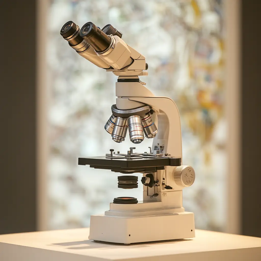
The Fascinating World of Microscopy: Viewing Mushroom Spores. Microscopy plays a crucial role in the field of mycology, especially when it comes to studying mushroom spores. Whether you’re a seasoned mycologist or a curious hobbyist, understanding how to view and analyze spores can open up a new dimension of knowledge and discovery. In this blog, we’ll explore the basics of microscopy for viewing mushroom spores, including how it relates to spore syringes and psilocybin spores.

Mushroom spores are the reproductive units of fungi, and they carry the genetic material necessary for the growth of new mushrooms. Studying these spores under a microscope allows mycologists to identify different species, observe spore morphology, and even determine the health of a spore syringe. For those interested in cultivating mushrooms, particularly psilocybin spores, microscopy provides valuable insights that can enhance both research and cultivation practices.
Spore syringes are commonly used in mushroom cultivation, and microscopy can help ensure the quality and purity of the spores within the syringe. By examining spores from a spore syringe under a microscope, you can check for contamination, verify the species, and assess the overall health of the spores. This step is especially important when working with psilocybin spores, as maintaining purity is critical for successful cultivation.
Psilocybin spores have gained significant attention in recent years due to the growing interest in psilocybin’s therapeutic potential. Researchers are using microscopy to study psilocybin spores, gaining insights into their genetic makeup and understanding how they develop into mushrooms. This research is not only advancing the field of mycology but also contributing to the broader understanding of psilocybin’s effects and applications.
Microscopy offers a fascinating window into the world of mushroom spores, providing valuable insights for both scientific research and mushroom cultivation. Whether you’re using a spore syringe to grow mushrooms or studying psilocybin spores for research, mastering the art of microscopy can greatly enhance your understanding and success. By following the steps outlined in this blog, you can begin exploring the intricate details of mushroom spores and contribute to the ever-growing field of mycology.
Remember, the world of mushroom spores is vast and full of discoveries waiting to be made—so grab your microscope and start exploring!